Procedures
Treacher Collins Syndrome (TCS) affects one in every 20,000 children in the U.S. each year. While this condition does not affect intellect, it impacts the development of the bones and other tissues of the face. There is no cure, but symptoms can be managed with surgical treatment and other therapies. Signs and symptoms will vary greatly, ranging from nearly imperceptible to severe. Most children who suffer from this condition have underdeveloped facial bones, especially the cheek bones, and a small chin and jaw (micrognathia). In some cases, the airway can be compromised by the small lower jaw, leading to serious, life-threatening health concerns and a tracheostomy at birth. Dr. Charles Thorne works with a multi-disciplinary team in New York, NY, performing a range of treatments to help manage the symptoms of TCS and improve your child's health and appearance. Once a member of a family develops TCS, 50 percent of that patient's children will also be affected.

Genetic mutations can cause Treacher Collins syndrome, which affects one in every 20,000 children in the U.S. each year.
Causes and Effects
TCS is caused by mutations that occur in specific genes during pregnancy. These mutations most commonly occur in the TCOF1 gene, but can also affect POLR1C or POLR1D genes. When the TCOF1 gene becomes mutated, TCS can become a dominant trait that can more easily be passed onto children. Mutations of the POLR1C or POLR1D genes will lead to recessive traits. As a result, 40 percent of children with TCS have inherited the condition from their parents. All other cases result from new mutations.
The effects of TCS can range from nearly imperceptible asymmetry to severe deformities in the face and head. Primarily, the condition is associated with an underdeveloped lower jaw. Micrognathia can result in difficulties with speech, eating, and breathing. Underdeveloped bones in the ear can lead to significant hearing issues, and malformed eyelids may affect the eyes as well. Many children can suffer from life-threatening disabilities that require tracheostomies and other serious interventions to manage the condition and its effects.
Dr. Thorne works with a multi-disciplinary team at Lenox Hill Hospital. Together, they can provide necessary treatment to manage TCS and improve the quality of life for your child.
Treatments and Therapies
Patients typically undergo multiple surgeries and treatments to manage the symptoms of TCS, including:
- Surgery to repair cleft palate and lip, if present
- Hearing aids for conductive hearing loss
- Bone and fat grafts to rebuild and augment underdeveloped cheeks
- Surgeries to address lower eyelid problems
- External ear reconstruction, which may include techniques for microtia such as grafts from rib cartilage
- Advancement of lower jaw to improve breathing and aesthetics
- Nose reconstruction
- Orthodontics
- Further fat or other tissue grafts to enhance features as the patient matures
Dr. Thorne works with a multi-disciplinary team at Lenox Hill Hospital. Together, they can provide necessary treatment to manage TCS and improve the quality of life for your child.
Contact Us Today
If your child has been diagnosed with Treacher Collins syndrome, contact our office online or call (212) 794-0044 to schedule a consultation with Dr. Thorne. He can perform a thorough evaluation to assess your child’s needs and develop an appropriate treatment plan.
Hemifacial microsomia, also known as craniofacial microsomia, is a congenital disorder that causes either one or both sides of the face to be underdeveloped. Hemifacial microsomia is the second most common congenital disorder affecting the face after cleft lip and palate. This condition most commonly affects one side of the face, more commonly the right side, and can involve the ear, soft tissue fulness, facial nerve function and the jaw bones. The condition may be mild with almost imperceptible asymmetry or severe with obvious asymmetry. In the most severe cases, patients with hemifacial microsomia have problems breathing and eating and may require a tracheostomy at birth. Patients with so called “isolated microtia” probably have the mildest form of hemifacial microsomia with a symmetrical or near-symmetrical face and an absent ear. Dr. Charles Thorne can perform surgical treatment for patients with hemifacial microsomia at his New York City practice.
“Dr. Charles Thorne and his team at the Lenox Hill Hospital can perform a variety of surgical treatments to improve facial symmetry, achieve normal occlusion and TMJ function, and enhance hearing.”

Severe hemifacial microsomia with underdevelopment of the right ear, soft tissue of the right cheek, facial nerve and the jaw bones on the right side.
Hemifacial Microsomia Causes
There is no known cause of craniofacial microsomia. We know that it occurs very early in pregnancy. We also know that it can be produced in animals by administering certain medications but these are not medications that humans would ever be prescribed. There has been no link to any specific medications or activities of the mother during pregnancy. The condition is generally NOT inherited but there are a few families around the world with more than one affected child which leads one to believe that there must be an inherited tendency or an inherited form of the condition.
During embryologic development, structures called the first and second brachial arches develop into the tissues of the face. In a healthy pregnancy, these structures will later develop into the jaws, the bone structure of the middle ear, the external ear, and the muscles that are used for chewing and facial expressions.
However, individuals with hemifacial microsomia experience a disruption in the formation of these facial features. Like microtia, hemifacial microsomia is tremendously variable. Some patients have facial asymmetry that is barely noticeable and others have severe asymmetry with absence of the ear, underdevelopment of the jaws. Underdevelopment of the soft tissue of the cheek and paralysis of some branches of the facial nerve. In fact, patients with microtia probably have hemifacial microsomia; it is just so mild that the facial asymmetry is not identified.
Treatments and Therapies
Hemifacial microsomia causes a wide range of difficulties. The patients who are severely affected require:
- Distraction of the short mandible early in childhood
- Fat grafting to the cheek or micosurgical reconstruction of the deficient cheek tissue
- Ear reconstructions
- Final jaw surgery
- Touchup fat grafting to the face
Dr. Thorne's team at the Lenox Hill Hospital can provide multi-disciplinary care for all stages of treatment.
Schedule Your Consultation
Dr. Charles Thorne has extensive experience helping patients improve their hearing and their appearance, and he wants to help you and your family. To learn more, or to schedule a personal consultation, contact our office today.
Ear deformities, also known as ear malformations or ear anomalies, include a wide range of birth defects that ultimately cause ears to be misshapen, deformed, or absent at birth. Dr. Charles Thorne has over 25 years of experience in the treatment of ear deformities, and can reconstruct a new ear for patients at his New York City practice. Dr. Thorne has provided considerable success for patients with microtia, Stahl’s ear, hemifacial microsomia, cryptotia, question mark ears, prominent ears, and macrotia, as well as post-traumatic deformities and several other conditions.

Dr. Thorne creating an ear framework from the patient’s rib cartilage for reconstruction of microtia.
Common Ear Deformities
Microtia can affect one or both ears, and is categorized into four different types, grades one through four.
Microtia: A congenital birth defect, microtia causes one or both ears to be malformed or completely absent. These defects can, and usually do, cause impaired hearing as well as self-consciousness associated with the appearance. Microtia most commonly appears in boys and is more common on the right side but can occur in either sex and on either side. As mentioned in the section on “Microtia” above, there is tremendous variation in the appearance of microtia. There are a number of classifications used to describe the severity of microtia but, from a practical point of view, the ear either looks acceptable to the patient or it doesn’t and if it doesn’t, then Dr. Thorne can help by reconstructing the auricle.
Hemifacial Microsomia: This condition is characterized by underdevelopment of the tissues on one, or rarely both, sides of the face. The ear, jaw bones, soft tissue and nerves can be affected. In some patients the asymmetry is barely noticeable and in others it is severe. The cause of hemifacial mircosomia is unknown. The sequence of treatment is based on the severity of the asymmetry. Dr. Thorne examines the patient with his team of physicians and specialists and, in consultation with the patient and family, the treatment plan is developed.
Stahl’s Ear: This condition involves the development of an extra ridge of cartilage, and can produce a pointed or "Dr. Spock" appearance of the ear. Correction can be performed at any age after 4 years.
Treatment for Ear Deformities
During your consultation, Dr. Thorne will determine which course of reconstruction is best for you or your child. Possible treatments may include:
Rib Cartilage Ear Reconstruction: Since the normal ear consists of skin overlying a flexible cartilage framework, it makes sense to use cartilage when reconstructing an ear. As shown in the image above, cartilage from the rib cage is spliced together to create a realistic framework. The framework is constructed do that it is the same size as the existing ear on the other side and is placed in a skin pocket, taking care that the position and angle are correct.
Medpor Framework and Prosthetic Ear Reconstruction: One of the alternatives to using the patient’s own cartilage is to use an artificial framework composed of polyethylene, known as Medpor. The procedure has the advantage of avoiding the scar and possible indentation on the chest, but the material is a hard, plastic substance and is not flexible. In addition, if the framework is exposed due to trauma from sports or other activities, it may not heal the way an ear made from the patient’s own tissues would heal. In general, Dr. Thorne believes that if a surgeon can carve a realistic framework from the patient’s cartilage, then that is a better option than using a foreign material.
Prosthetic Ear Reconstruction: Prosthetic reconstruction involves creation of an entirely artificial ear as one might see in a wax museum. The artificial ear is attached to the side of the head with glue or with titanium implants much like those used to attach dentures. Prosthetic ears can be made very realistic but most patients, particularly young ones, will not wear them. Prostheses are a daily reminder that something is wrong rather than a solution that allows the patient to move on. Prosthetic reconstruction can be very helpful for older patients who have lost an ear or part of an ear from skin cancer and would like a prosthesis for social events. The other disadvantage of prosthetic ears is that they have to be replaced every few years for the life of the patient.
Schedule Your Consultation
Dr. Thorne understands the impact that ear deformities can have on a patient and a family. It is his mission to provide state-of-the-art surgical care for patients with ear malformations and he has been doing so for three decades.
Dr. Thorne also stays at the cutting edge of technology research and would be happy to present to you the current state of science and the future possibilities of bioprinting an ear framework from the patient’s own ear cartilage. This process is not available now but may be in the future.
Contact our office to schedule your personal consultation.
Most patients with microtia also have aural atresia, which means absence of an ear canal. Patients who do not have an ear canal may be able restore their hearing with aural atresia repair or by use of a Bone Anchored Hearing Aid (BAHA). Dr. Charles Thorne in Lenox Hill, Manhattan, partners with a talented and trusted otologist for rebuilding the inner ear. A BAHA can be worn immediately after birth. Aural atresia repair is best performed after the auricular reconstruction.
Understanding Aural Atresia Repair and BAHAs
Aural atresia repair involves restoring inner ear hearing loss through the surgical construction of an ear canal and an ear drum. A Bone Anchored Hearing Aid on the other hand consists of two types:
- Soft Band BAHA: During this procedure, a small processor is attached to a band worn around the head that receives and sends sounds to the middle ear bones, transmitting sound waves to the brain. This is traditionally chosen for infants with middle ear or conductive hearing loss.
- BAHA (Bone Anchored Hearing Aid): Rather than remaining on a band around the head, the processor is connected to a titanium post in the skull. Patients must be at least five years old and have adequate skull density to undergo this procedure. This type of aid provides the best possible replacement for the middle ear function.
To determine which option is most appropriate, Dr. Thorne will conduct a thorough assessment of you or your child's ears during a consultation.


This patient underwent both an aural atresia repair and a reconstruction of the external ear.
What are the Benefits?
Patients who undergo aural atresia repair or who wear a BAHA can enjoy restored hearing almost immediately. This is especially beneficial for young patients who have aural atresia on both sides and are functionally deaf without hearing aids or aural atresia repair. When this procedure is combined with rib cartilage ear reconstructive surgery, patients can have both their hearing and outer ear aesthetics restored, providing a better quality of life and improved self-confidence.
Who is a Candidate?
Unfortunately, aural atresia repair is not always a viable option. About 50 percent of patients with microtia are candidates for aural atresia repair based on the extent of the deformity of the middle ear structures. Patients with Treacher Collins syndrome are almost never candidates for surgical repair of the aural atresia. Fortunately, almost all patients with microtia are candidates for the BAHA which actually provides superior hearing in the long term compared to aural atresia repair.
What to Expect During the Procedure
As one of the more challenging ear reconstruction surgeries available, aural atresia repair is always performed by an experienced otologist. A typical procedure usually includes providing the patient with a sterile, skin-lined external auditory canal in addition to improving hearing. Dr. Thorne works closely with a trusted and skilled otologist to make sure patients achieve optimal results.
Patients who undergo aural atresia repair can enjoy restored hearing almost immediately
If your child suffers from congenital aural atresia in addition to microtia, we encourage you to make an appointment with Dr. Thorne to determine if he or she is a candidate for aural atresia repair or a BAHA. The sooner treatment can be performed, the greater their chance of having hearing restored during crucial developmental stages.
Learn if Aural Atresia Repair is Right for You
Please contact our office today online or give us a call at 212-794-0044 to schedule your consultation with Dr. Thorne. He will perform a complete examination and outline the most suitable treatment options.
Related Articles
START YOUR INQUIRY!
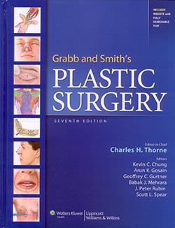
Dr. Thorne is the Editor-in-Chief and the author of several chapters in Grabb and Smith's PLASTIC SURGERY, 7th Edition.
Ear Construction Chapter in PDF


